
Arraystar GlycoRNA Array Profiling
Arraystar GlycoRNA Array combines methods of biochemical capture of glycoRNA and RNA detection by microarray to quantify and profile glycoRNA expression. The integration of these two advanced techniques leverages the strengths of both methods for high specificity, sensitivity, and accuracy.
The array covers a wide range of glycosylated small RNA classes, including Y-RNAs/Y-RNA fragments, tRNAs, tsRNAs (tiRNAs & tRFs), pre-miRNAs, miRNAs, snRNAs/snRNA fragments, snoRNAs/snoRNA fragments, rRNAs/rRNA fragments, and scRNAs.
Using this cutting-edge approach, researchers can gain comprehensive glycoRNA expression details to discover and understand this new class of RNA molecules in gene regulation, cellular functions and human diseases.
GlycoRNA Detection Methods
GlycoRNAs can be detected, captured, and enriched from the total RNA samples by highly specific biochemical methods to ensure genuine glycoRNA signals and increased the detection sensitivity.
• Metabolic Labeling and Click Chemistry
GlycoRNAs are metabolically labeled with the use of click chemistry [1](Fig.1). The method utilizes reporter sugars, such as N-azidoacetylmannosamine (Ac4ManNAz) with an azide group, to be incorporated into the glycans in the cells or tissues. The glycan of isolated RNA is then conjugated between the azide and a label such as dibenzocyclooctyne-biotin (DBCO-biotin) with azide-alkyne cycloaddition click chemistry (Fig.1).
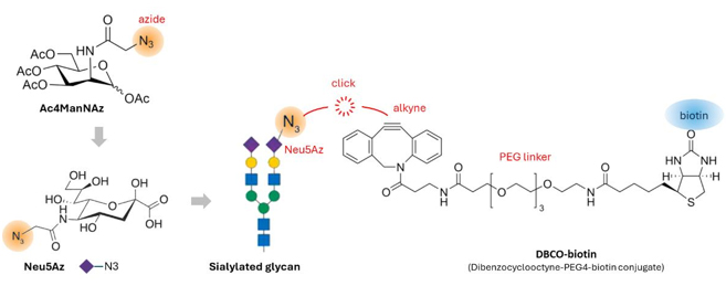
Fig.1 Ac4ManNAz (N-azidoacetylmannosamine-tetraacylated) is a precursor for Neu5 sialic acid (N-Acetylneuraminic acid). It is peracetylated to increase cell permeability during cell culture. In cells, Ac4ManNAz is metabolically converted to Neu5Az to become part of the sialylated glycan. In vitro, the azide in Neu5Az and the alkyne in the DBCO-biotin click reagent are linked by copper-free click chemistry, allowing the glycan to be labeled with biotin.
• Periodate Oxidation and Aldehyde Ligation (pAL)
The periodate oxidation and aldehyde ligation method (pAL) is designed to label native glycoRNAs without the need for metabolic labeling in living cells [2]. This method uses periodate to selectively oxidize the sialic acid diols of the glycoRNA into aldehyde group under optimized condition, followed by aldehyde ligation with the amine group of aldehyde reactive biotin reagent (Fig. 2). rPAL is robust and flexible, with sensitivity at least an order of magnitude higher than previous methods for detecting sialoglycoRNAs.
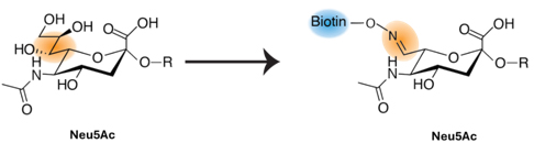
Fig.2 In RNA Periodate oxidation and Aldehyde Ligation (rPAL), the sialic acid diols can be oxidized to aldehyde by periodate, and then ligated to the amine group of the aldehyde reactive label such as biotin.
• In Vivo Cell Periodate Oxidation and Aldehyde Labeling (pAL)
In vivo Cell Periodate oxidation and aldehyde labeling (pAL) of glycoRNA is used to label glycoRNAs on living cell surfaces and in cell membrane-associated RNAs with fluorescent dye or biotin [3]. Specifically, Cy5-hydrazide or biotin-hydrazide is conjugated to the cell surface glycoRNAs treated with mild oxidation using sodium periodate.
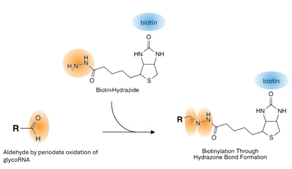
Fig.3 Sialic acid diols in glycoRNA are oxidized with periodate to form aldehyde group. Biotin- or Cy5- hydrazide is then conjugated to the glycoRNA via the aldehyde group.
• Cell Surface Imaging of GlycoRNAs
The glycoRNA expression and localization on cell surface can be visually imaged, for example, by sialic acid aptamer and RNA in situ hybridization-mediated proximity ligation assay (ARPLA) [4]. As shown in Fig. 4, ARPLA consists of the following functional components: (1) a glycan probe, with an ‘aptamer’ to selectively bind N-acetylneuraminic acid (Neu5Ac), a ‘spacer’ to avoid steric hindrance during hybridization, and a DNA linker (‘linker G’) for subsequent proximity ligation; (2) an RNA-binding probe with a DNA strand for RNA in situ hybridization “RISH”, another ‘spacer’ and a DNA linker (‘linker R’) that works with linker G; (3) connectors 1 and 2 that hybridize with linkers G and R to allow in situ ligation to generate circular DNA as RCA template, and (4) a reporter composed of fluorophore-conjugated single-stranded DNA (ssDNA) probes complementary to the RCA product to report the glycoRNA detection signals. The dual glycan-RNA binding and in situ ligation ensure high selectivity and low false positives. The rolling circle amplification generates highly sensitive signals.
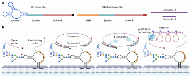
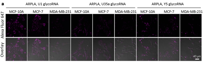
Fig.4 GlycoRNA imaging using ARPLA. (a) Schematic presentation of the glycan probe, RNA-binding probe, and connectors. (b) Their simultaneous binding to the glycoRNA allows T4 DNA ligase ligation to occur to form template for rolling circle amplification (RCA) using phi29 DNA polymerase. The amplified RCA products are detected by fluorescent reporter probe hybridization. (c) ARPLA imaging of U1, U35a and Y5 glycoRNAs in MCF-10A, MCF-7 and MDA-MB-231 cells.
• Lectin Affinity Binding
Lectins, e.g. wheat germ agglutinin (WGA) and Maackia amurensis Lectin II (MALII), recognize and bind directly to sialylated glycan structures present on glycoRNAs [1]. GlycoRNA purification based on biotin-lectin affinity binding is an effective way to isolate and enrich glycosylated RNAs.
Bioinformatics Tools
GlycoRNA studies can be greatly aided by bioinformatics tools. For example, GlyinsRNA can predict putative glycosylated sites on RNA molecules [5]. It uses glycosylated and un-glycosylated small RNA sequences [6] to train machine learning models. GlyinsRNA is implemented as an online webserver, where both the predicted glycosylation sites and the overrepresented RNA-binding protein (RBP)-related motifs are annotated.
Functional Studies
• GlycoRNA Binding Proteins
The glycoRNA bound partner proteins provide excellent indications of what functions the glycoRNAs do. The interacting proteins can be either bind the glycan (e.g. siglecs) or RNA moiety (RBPs). For example, to investigate the immunomodulatory potential of glycoRNAs, the interactions between glycoRNAs and members of the sialic acid-binding immunoglobulin-like lectin (Siglec) receptor family can be evaluated by immunological assays [1, 7]. By examining these interactions, scientists can gain insights into how glycoRNAs may influence immune responses and contribute to various disease processes, particularly in the context of neuroinflammation and autoimmune disorders [1, 7]. Conversely, glycoRNAs can be screened as ligands for many orphan receptors with unknown cognate ligands.
• Genetic, Pharmacological, Enzymatic Approaches
Both genetic and pharmacological inhibition approaches have been used to study the biosynthesis and functions of glycoRNAs. For example, genetic ldlD mutant cell line lacking the ability to interconvert UDP-glucose(Glc)/GlcNAc into UDPgalactose, CRISPR-Cas9 knockout of UDP-galactose-4-epimerase, and glycan-biosynthesis pharmacological inhibitors (e.g. NGI-1, Kifunensine, Swainsonine) have been used to observe glycan biosynthetic machinery on glycoRNA biogenesis [1]. A panel of endoglycosidases with different specificities for glycan structures, e.g. sialidase, PNGaseF, Endo-F2, -F3, -Hf, and O-deglycosidase, can be used to dissect the glycan structure and the glycoRNA functions [1]. Such studies have provided valuable insights into the mechanisms underlying glycoRNA formation and their potential roles in various cellular processes and disease states.
Related Service
Related Reviews
References
1. Flynn, R.A., et al. (2021) “Small RNAs are modified with N-glycans and displayed on the surface of living cells” Cell 184(12):3109-3124 e22 [PMID:34004145]
2. Hemberger, H., et al. (2023) “Rapid and sensitive detection of native glycoRNAs” bioRxiv 2023.02.26.530106
3. Abledu, J.K., et al. (2024) “Cell surface RNA expression modulates alveolar epithelial function” bioRxiv 2024.05.19.594844
4. Ma, Y., et al. (2024) “Spatial imaging of glycoRNA in single cells with ARPLA” Nat Biotechnol 42(4):608-616 [PMID:37217750]
5. Cui, C., et al. (2021) “GlyinsRNA: a webserver for predicting glycosylation sites on small RNAs” RNA Biol 18(sup2):600-603 [PMID:34559595]
6. Flynn, R.A., et al. (2019) “Mammalian Y RNAs are modified at discrete guanosine residues with N-glycans” bioRxiv 787614
7. Zhang, N., et al. (2024) “Cell surface RNAs control neutrophil recruitment” Cell 187(4):846-860 e17 [PMID:38262409]